The combination of CBCT scans with zygomatic dental implants has transformed the way advanced implant dentistry is planned and performed. Especially in patients with severe upper jaw bone loss, CBCT imaging provides the three-dimensional precision needed to safely place implants into the dense zygomatic (cheek) bone. At MaxFax Zygoma Center, led by Prof. Dr. Celal Çandırlı, CBCT scans with zygomatic dental implants are an essential part of diagnosis, surgical planning, risk management, and long-term follow-up. Understanding how CBCT technology works and why it matters helps patients feel more confident when choosing this life-changing treatment.
What Are CBCT Scans?
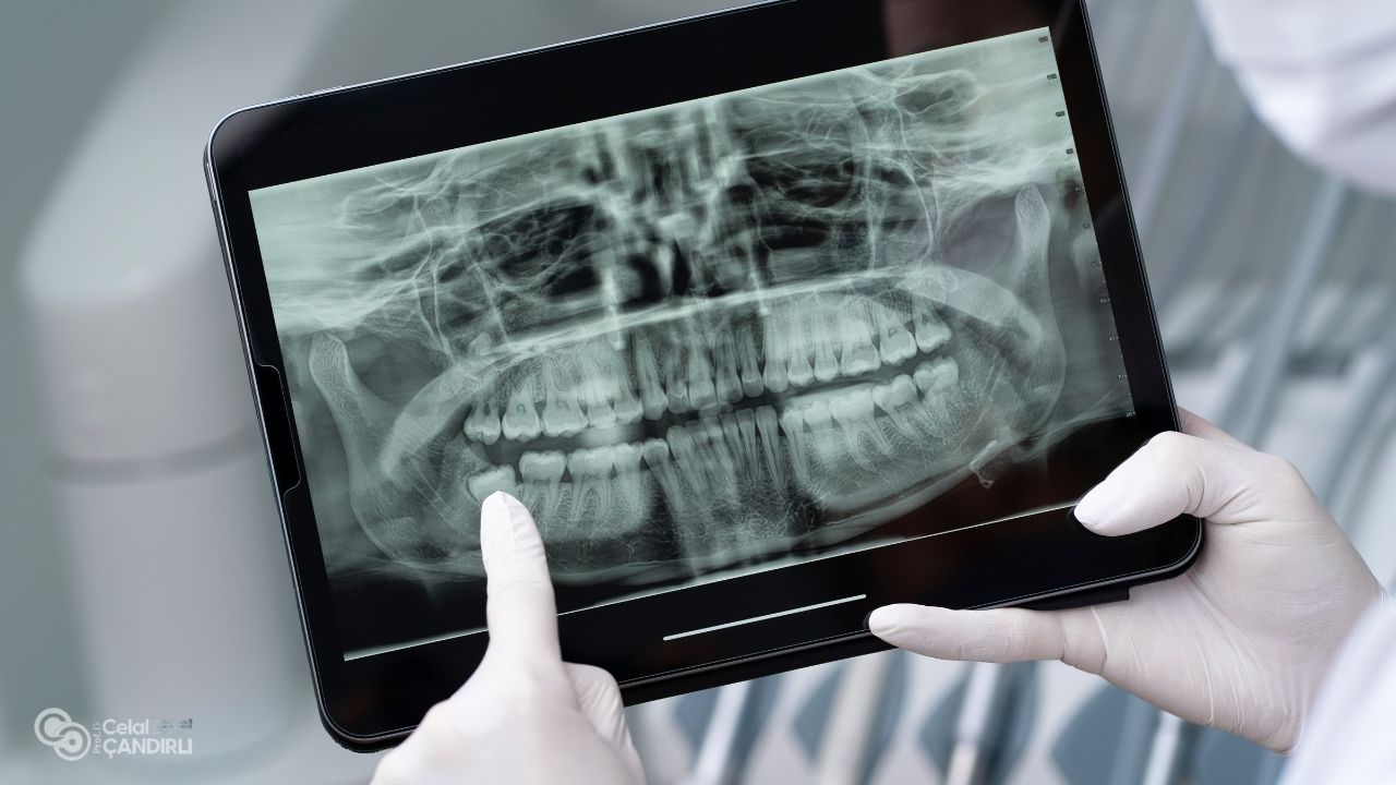
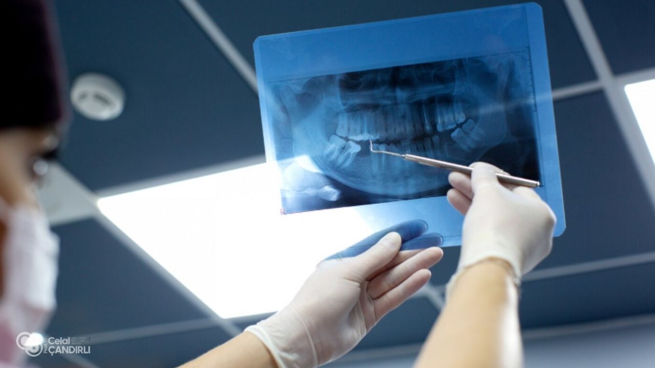
CBCT stands for Cone Beam Computed Tomography. Unlike traditional two-dimensional panoramic x-rays, CBCT provides detailed three-dimensional images of the jaw, sinuses, teeth, zygomatic bone, and surrounding anatomical structures. This level of detail is crucial when performing CBCT scans with zygomatic dental implants because the surgical field lies close to delicate structures such as the sinuses, orbital floor, and nerves. The clarity and accuracy of CBCT images make it possible to create a safe and predictable surgical plan tailored to each patient’s anatomy.
Why CBCT Is Superior to Traditional X-Rays
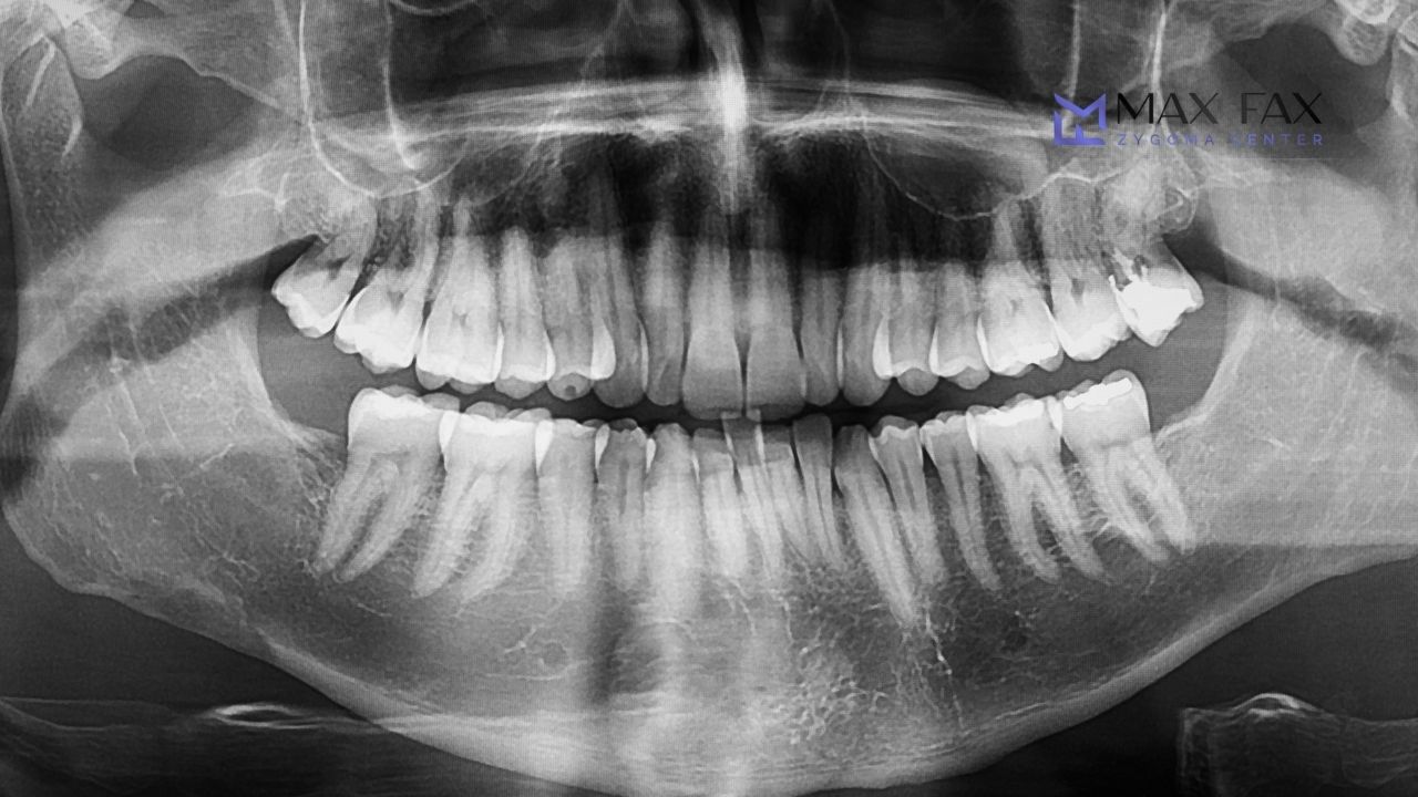
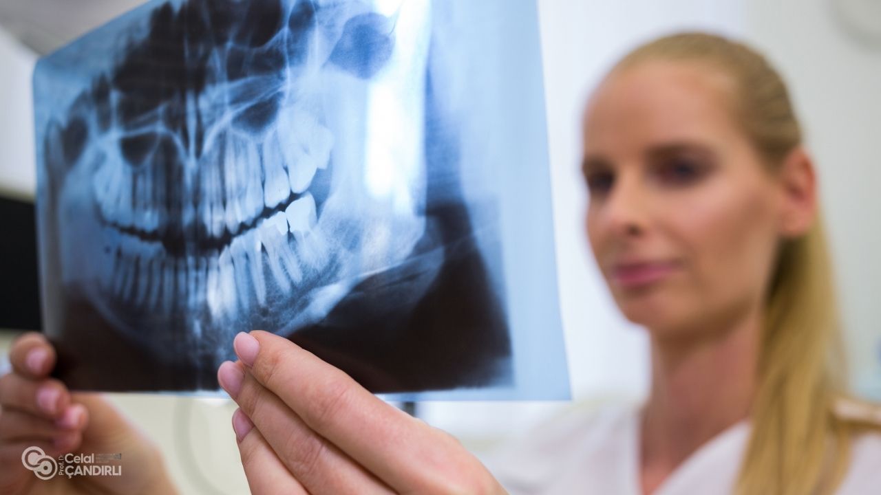
Traditional dental x-rays give only flat, two-dimensional information. For simple dental procedures this may be enough, but for complex maxillary rehabilitation it is insufficient. With CBCT scans with zygomatic dental implants, clinicians can rotate and analyze the scanned area from every angle. This allows them to evaluate bone density, detect sinus conditions, identify exact nerve positioning, and map ideal implant trajectories. Because zygomatic implants are much longer and placed at steep angles, accurate visualization is essential to avoid complications and ensure implant stability.
Why CBCT Scans Are Essential for Zygomatic Dental Implants?
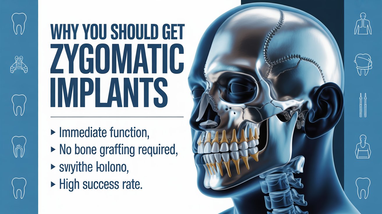
Zygomatic dental implants are used when the upper jawbone has resorbed to the point where conventional implants cannot be placed. Instead of anchoring in the weakened maxilla, these implants extend into the zygomatic bone. The proximity to the sinus and orbital structures means that CBCT scans with zygomatic dental implants are not optional; they are a fundamental requirement for safe and successful treatment. CBCT enables the surgeon to assess the location and shape of the sinus cavity, measure the thickness and quality of the zygomatic bone, and choose the ideal implant length and angle.
The Role of CBCT in Treatment Planning
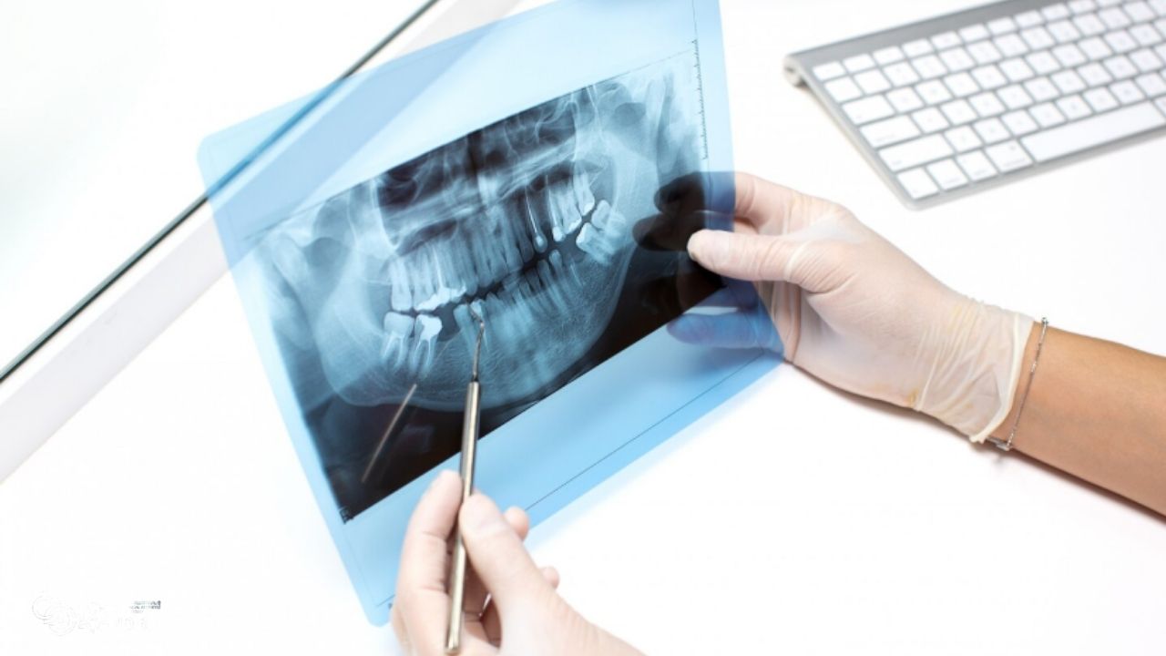
Before surgery, the CBCT data is imported into advanced digital planning software. This allows the surgeon to virtually position implants and simulate the procedure. In CBCT scans with zygomatic dental implants, every millimeter counts. Virtual planning ensures that each implant enters and exits the bone at exactly the right point, avoiding nerves and vital spaces. This planning stage supports greater precision during surgery and reduces unnecessary guesswork.
How CBCT Scans Improve Surgical Safety
Safety is one of the greatest advantages of using CBCT scans with zygomatic dental implants. Because these implants are placed near the sinus and beneath the eye socket, misjudgment in angulation or depth can lead to complications. CBCT scans provide the surgeon with accurate three-dimensional knowledge of the patient’s anatomy so critical structures can be clearly avoided. The technology also reveals any existing sinus disease, cysts, or anatomical variations that may alter the surgical approach.
Risk Reduction Through Advanced Imaging
By identifying anatomical risks in advance, CBCT scans with zygomatic dental implants significantly reduce the likelihood of perforation, nerve involvement, implant misplacement, or sinus complications. This allows the surgeon to enter the operating room with a clearly defined, patient-specific plan. The result is greater predictability, shorter surgical times, and higher success rates.
CBCT Scans and Guided Surgical Techniques
In many cases, digital data from CBCT scans with zygomatic dental implants is also used to design surgical guides. These guides support precise drill positioning according to the virtual plan. Guided surgery improves accuracy and consistency, especially in complex cases with little remaining bone. This level of control is particularly important in severe maxillary atrophy where zygomatic implants may be the only fixed solution available.
Benefits for Immediate Loading Protocols
Many patients receiving zygomatic implants benefit from immediate loading techniques, meaning teeth are placed within a very short time after surgery. CBCT scans with zygomatic dental implants support this process by confirming ideal implant stability and positioning. When implants are placed in dense zygomatic bone using digitally planned trajectories, they achieve strong primary stability, making immediate functional restoration possible in appropriate cases.
How CBCT Helps Customize Prosthetic Design
Zygomatic implants are only one part of rehabilitation. The prosthetic teeth must also be designed to function harmoniously with the jaw and muscles. CBCT scans with zygomatic dental implants allow prosthodontists to study the available restorative space, lip support, occlusion patterns, and skeletal relationships. This helps ensure the final prosthesis is both comfortable and aesthetically natural.
Improved Communication with Patients
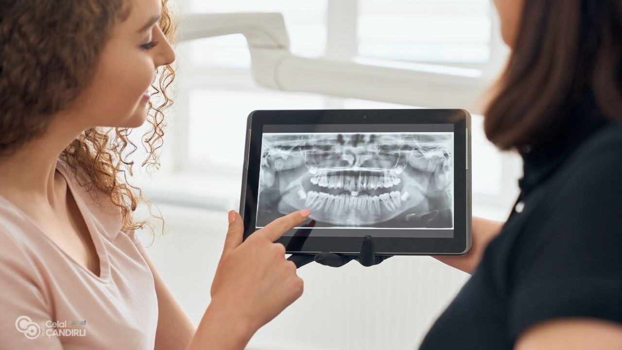
CBCT scans with zygomatic dental implants are also an important educational tool. Patients can clearly see the condition of their jawbone, the limitations of traditional implants, and why zygomatic implants are recommended. Viewing their anatomy in 3D helps patients understand the steps of treatment, reducing anxiety and increasing trust in the process.
Post-Operative Evaluation Using CBCT Scans
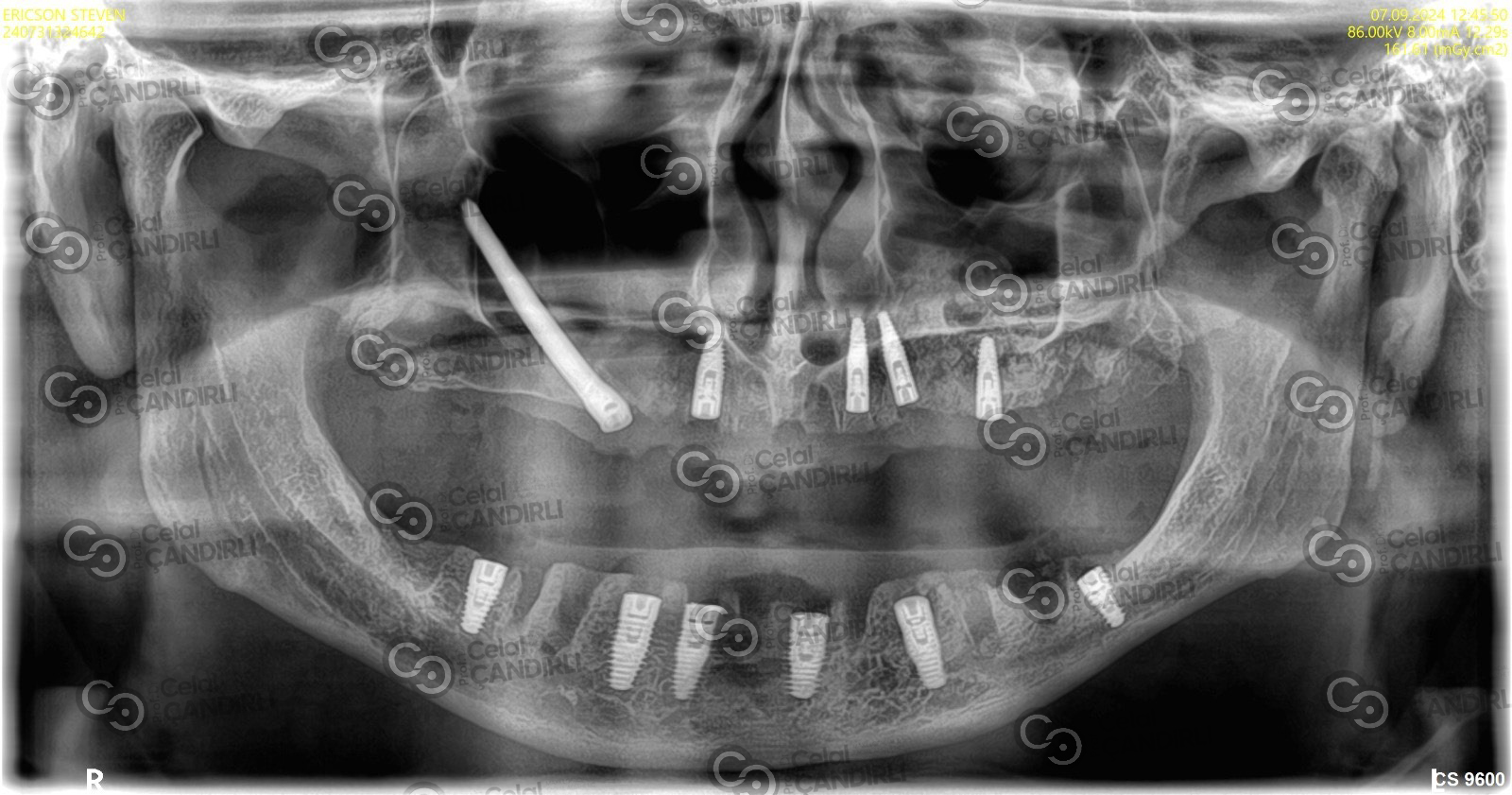
After surgery, CBCT scans with zygomatic dental implants may also be used for follow-up assessment. Post-operative CBCT images confirm implant position, integration with bone, and sinus health. This creates a reliable baseline for long-term monitoring. Any changes can be detected early and managed promptly to protect the success of the treatment.
CBCT Radiation Safety Considerations
Many patients naturally ask about radiation exposure. CBCT scans with zygomatic dental implants use significantly lower radiation than medical CT scans. Modern CBCT machines are designed specifically for dental and maxillofacial use, balancing image quality with safety. The diagnostic value gained from CBCT far outweighs the minimal exposure when imaging is performed responsibly under professional care.
The Future of CBCT in Implant Dentistry
Digital dentistry continues to evolve rapidly. CBCT scans with zygomatic dental implants are now being integrated with artificial intelligence, digital impression systems, and 3D printing. These technologies allow fully customized treatment plans and prostheses made uniquely for each patient. The combination of CBCT imaging and zygomatic implant techniques will continue to set the standard for treating severe bone loss without the need for extensive grafting.
Why Choose Prof. Dr. Celal Çandırlı
At MaxFax Zygoma Center, Prof. Dr. Celal Çandırlı uses CBCT scans with zygomatic dental implants as a cornerstone of treatment planning and execution. His expertise ensures that every stage of care is based on precise anatomical knowledge and evidence-based protocols. Patients benefit from individualized treatment strategies, technologically advanced care, and compassionate clinical support from the first consultation through long-term follow-up.
Frequently Asked Questions About CBCT Scans with Zygomatic Dental Implants
The use of CBCT scans with zygomatic dental implants has become essential in modern implant dentistry. This technology allows surgeons to visualize complex anatomy, plan implant placement with accuracy, reduce risk, and achieve stable, long-lasting outcomes. For patients with severe upper jaw bone loss, CBCT-guided zygomatic implants provide a powerful alternative to traditional bone grafting procedures. Under the expertise of specialists such as Prof. Dr. Celal Çandırlı at MaxFax Zygoma Center, patients receive treatment that combines advanced digital imaging with surgical excellence, restoring both function and confidence through safe and predictable implant rehabilitation.
What does a CBCT scan show?
A CBCT (Cone Beam Computed Tomography) scan shows a three-dimensional image of the teeth, jaws, sinuses, airway, temporomandibular joints, and surrounding bone and soft tissues. Unlike traditional dental X-rays, CBCT provides detailed cross-sectional views, allowing dentists and surgeons to assess bone volume, density, tooth position, impacted teeth, pathology, and nerve locations with high precision. This level of detail is especially valuable for implant planning, orthodontics, jaw surgery, sinus evaluation, and diagnosing complex dental problems.
What are the 3 views in CBCT?
CBCT imaging typically provides three main anatomical views: axial, coronal, and sagittal. The axial view slices the head horizontally, the coronal view slices it vertically from front to back, and the sagittal view slices it vertically from left to right. Together, these three planes allow clinicians to examine structures from multiple angles, making diagnosis and treatment planning much more accurate compared to 2D imaging.
Are CBCT and CT scan the same?
CBCT and medical CT scans are similar in principle, but they are not the same. A medical CT uses a fan-shaped beam and multiple rotations to capture soft tissues and organs in great detail, typically with higher radiation. A CBCT scan uses a cone-shaped beam and one rotation to focus mainly on hard tissues such as teeth and bones, with significantly lower radiation and cost. CBCT is designed specifically for dental and maxillofacial imaging.
What does a CBCT scan cost?
The cost of a CBCT scan varies depending on the country, clinic, and complexity of the scan. Generally, it ranges from the equivalent of a few hundred to several hundred dollars or euros. Full-arch scans used for implant planning typically cost more than localized scans. Although it is an added diagnostic expense, CBCT often prevents complications and improves treatment accuracy, making it a worthwhile investment in many cases.
What are the risks of CBCT?
The primary risk of CBCT is exposure to radiation, although the dose is much lower than that of a medical CT scan. Modern machines use optimized low-dose settings to minimize exposure. Other risks are minimal, but unnecessary repeat scans should be avoided. Pregnant patients should inform their clinician before imaging so that safety precautions can be taken or imaging postponed when appropriate.
What are the four types of scans?
In a broad medical context, the four commonly referenced scan types are X-rays, CT scans, CBCT scans, and MRI scans. X-rays provide 2D images mainly of bones and teeth. CT scans produce detailed 3D images of both soft and hard tissues. CBCT scans generate 3D images mainly of the jaws and facial structures with lower radiation than medical CT. MRI scans use magnetic fields to visualize soft tissues such as muscles, joints, and nerves without radiation exposure.
Can CBCT detect tumors?
Yes, CBCT can sometimes detect tumors, cysts, abnormal growths, and other pathological changes in the jaw and facial bones. However, CBCT is not always the primary diagnostic tool for soft tissue tumors. If a suspicious lesion is detected, further evaluation with medical CT, MRI, or biopsy may be recommended to confirm the diagnosis and assess its nature and extent.
What is the 80/20 rule in dentistry?
The 80/20 rule in dentistry, adapted from the Pareto principle, suggests that roughly 80% of dental problems arise from 20% of patients or 20% of risk behaviors. It highlights the importance of preventive care, patient education, and identifying high-risk groups. In practice, it reminds clinicians that focused attention on key contributing factors can greatly reduce overall disease burden.
Can CBCT show nerve damage?
CBCT scans can clearly show the position and course of nerves, especially the inferior alveolar nerve in the lower jaw. While CBCT does not always show actual nerve damage itself, it is extremely useful in identifying compression risks, trauma effects, or implant proximity to nerves. This helps clinicians plan safer surgeries and assess causes of numbness or altered sensation when they occur.